What is Ultrasound Imaging of the Musculoskeletal System?
Ultrasound imaging, also called ultrasound scanning or sonography, involves exposing part of the body to high-frequency sound waves to produce pictures of the inside of the body. Ultrasound exams do not use ionizing radiation (as used in x-rays). Because ultrasound images are captured in real-time, they can show the structure and movement of the body's internal organs, as well as blood flowing through blood vessels.
Ultrasound imaging is a noninvasive medical test that helps podiatrists diagnose and treat medical conditions.
Ultrasound images of the musculoskeletal system provide pictures of muscles, tendons, ligaments, joints and soft tissue throughout the body.
What are some common uses of the procedure?
Ultrasound images are typically used to help diagnose:
What does the equipment look like?
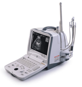 Ultrasound scanners consist of a console containing a computer and electronics, a video display screen and a transducer that is used to scan the body and blood vessels. The transducer is a small hand-held device that resembles a microphone, attached to the scanner by a cord.
The transducer sends out high frequency sound waves into the body and then listens for the returning echoes from the tissues in the body. The principles are similar to sonar used by boats and submarines.
Ultrasound scanners consist of a console containing a computer and electronics, a video display screen and a transducer that is used to scan the body and blood vessels. The transducer is a small hand-held device that resembles a microphone, attached to the scanner by a cord.
The transducer sends out high frequency sound waves into the body and then listens for the returning echoes from the tissues in the body. The principles are similar to sonar used by boats and submarines.
The ultrasound image is immediately visible on a nearby screen that looks much like a computer or television monitor. The image is created based on the amplitude (strength), frequency and time it takes for the sound signal to return from the patient to the transducer.
Ultrasound imaging is based on the same principles involved in the sonar used by bats, ships and fishermen. When a sound wave strikes an object, it bounces back, or echoes. By measuring these echo waves it is possible to determine how far away the object is and its size, shape, and consistency (whether the object is solid, filled with fluid, or both).
In an ultrasound examination, a transducer both sends the sound waves and records the echoing waves. When the transducer is pressed against the skin, it directs small pulses of inaudible, high-frequency sound waves into the body. As the sound waves bounce off of muscles, bones, fluids and tissues, the sensitive microphone in the transducer records tiny changes in the sound's pitch and direction. These signature waves are instantly measured and displayed by a computer, which in turn creates a real-time picture on the monitor. One or more frames of the moving pictures are typically captured as still images.
How is the procedure performed?
For most ultrasound exams of the musculoskeletal system, the patient is seated on an examination table. For some ultrasound exams, the patient is positioned lying face-up on an examination table that can be tilted or moved.
A clear water-based gel is applied to the area of the body being studied to help the transducer make secure contact with the body and eliminate air pockets between the transducer and the skin. The doctor then presses the transducer firmly against the skin and sweeps it over the area of interest.
When the examination is complete, the patient may be asked to dress and wait while the ultrasound images are reviewed. However, the doctors are often able to review the ultrasound images in real-time as they are acquired and the patient can be released immediately.
This ultrasound examination is usually completed within 15-30 minutes but may occasionally take longer.
What will I experience during and after the procedure?
Most ultrasound examinations are painless, fast and easy.
After you are positioned on the examination table, the doctors will apply some warm water-based gel on your skin and then place the transducer firmly against your body, moving it back and forth over the area of interest until the desired images are captured. There is usually no discomfort from pressure as the transducer is pressed against the area being examined.
If scanning is performed over an area of tenderness, you may feel pressure or minor pain from the transducer.
The doctor may ask you to move the extremity being examined or may move it for you to evaluate not only anatomy but also function of a joint, muscle, ligament or tendon.
Once the imaging is complete, the gel will be wiped off your skin.
What are the benefits vs. risks?
BenefitsWhat are the limitations of Ultrasound Imaging of the Musculoskeletal System?
Ultrasound has difficulty penetrating bone and therefore can only see the outer surface of bony structures and not what lies within. For visualizing internal structure of bones or certain joints, other imaging modalities such as MRI are typically used.